New brain scans may show if a concussion has not yet healed
Delta waves may help decide when it’s safe for teen athletes to return to play

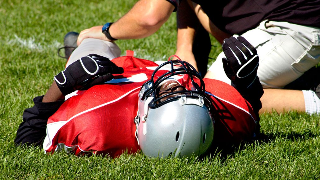
Football poses one of the highest concussion risks in youth sports. But many players with brain injuries hide their symptoms to return to play, risking long-term damage. Researchers hope to guard against that.
Serega/Getty Images
Share this:
- Share via email (Opens in new window) Email
- Click to share on Facebook (Opens in new window) Facebook
- Click to share on X (Opens in new window) X
- Click to share on Pinterest (Opens in new window) Pinterest
- Click to share on Reddit (Opens in new window) Reddit
- Share to Google Classroom (Opens in new window) Google Classroom
- Click to print (Opens in new window) Print
Admitting you’re in pain isn’t easy after a hard hit to the head in football. You may not want to let your team down, disappoint the coach or seem like a softie. But hiding your symptoms makes it hard to know if you have a concussion — and if so, when it’s safe for you to return to play. As an engineering student, Elizabeth Davenport set out to make those assessments easier.
Back in 2011, she recalls, “I wanted to give players, parents and doctors an objective number.” That is, she wanted some quantifiable rating of a head injury’s severity. That’s important when 78 percent of college athletes report hiding concussion symptoms.
Davenport is now a biomedical engineer at the University of Texas Southwestern Medical Center in Dallas. Her team thinks it may have just found such an injury rating system. It measures a type of brain wave. The number holds promise for diagnosing concussions. One drawback: It requires specialized equipment. That means it isn’t easy to obtain — yet.
Peering at players’ brains
Davenport is part of a team that recruited 128 high school boys between 2012 and 2019 to take part in the study. All were football players around 16 years old. Brain scans of 50 of these players were available at the start and end of a season, about four months apart. The researchers focused on a measure of their brain activity known as delta waves.
During football seasons, 11 of those 50 athletes suffered a concussion playing the sport. Researchers were able to scan the brains of eight of them within three days of their injury. The scientists compared delta waves of those eight boys with two control groups: eight non-concussed football players and eight boys about the same age who took part in noncontact sports, such as swimming and tennis.
Davenport’s team measured brain waves with a new technology. It’s known as MEG. That’s short for magnetoencephalography (Mag-NEE-toh-en-sef-uh-LAH-gruh-fee). Only 40 MEG scanners exist in U.S. clinics today. (Doctors mostly use them to plan epilepsy surgeries.) Like more common EEG brain wave scanners, MEG records the activity of neurons. These brain cells work by sending and receiving electromagnetic signals.
Both EEGs and MEGs display brain activity as waves of different frequencies. Delta waves have the lowest frequency, just 1 to 4 hertz (cycles per second).
Delta waves in players who sustained a concussion differed from those in the control groups. These waves therefore seem like a potential marker of concussions, the researchers say. They shared their findings in September in Brain and Behavior.
Why MEG and delta waves?
EEG scanners cannot detect weak electromagnetic signals deep inside the brain. These scanners attach only 20 to 30 electrodes to the scalp. MEG devices, in contrast, use 306 wireless sensors to record those signals anywhere in the brain’s folds and grooves.
Davenport also took magnetic resonance images (MRIs) of the players’ brains. Those images show the brain’s physical features, something like a satellite’s street view in Google Maps. MEG overlays the activity track on that street view. Doing both scans takes about 30 minutes. The final images capture the brain’s activity in much greater detail than EEG scans.
Researchers already knew from EEG scans that everyone has delta waves during deep sleep. These waves turn up in awake adults only after concussions or chronic brain damage from disease. But looking for delta wave changes due to injuries proved tricky in teen brains, even with the detailed MEG scans. That’s because all kids have delta waves while awake, injured or not. (Delta waves decrease with age as part of the brain’s normal remodeling.)
That made it much harder for the researchers to tell if delta wave changes in teen brains were due to recent concussions or normal development. Since every brain develops at a different rate, Davenport’s analysis relied on comparing a player’s preseason and postseason scans.
After their brain injuries, concussed teens’ average delta waves were higher than they had been at baseline. The averages were lower in the control groups, as expected with normal development.
What about real sidelines?
Getting a brain scan done within 72 hours of an injury did not prove to be easy, Davenport notes. In the end, her team could compare only eight concussed players to eight boys in each control group. To check if the findings hold up, she says, it will be important to repeat the analysis in more kids.
But even if the results are confirmed, the next challenge will be to use them in the real world, says Danielle Sandsmark. She’s a neurologist at the University of Pennsylvania in Philadelphia. The problem: Without preseason scans on every player, it’s not clear if delta waves can quickly diagnose concussions. And because MEG scanners are costly and not widely available, those preseason scans may not exist.
“It would be great if we could translate the [data] we need into a less cumbersome technology [than MEG],” says Sandsmark. She works on developing blood tests that could predict whose brains may heal more slowly. That’s about one in every five concussion patients. Right now, doctors can’t tell who would fall into that group. That makes return-to-play decisions in football hard. Finding chemical clues in blood tests that reflect delta wave changes would help.

Even better than a blood test would be a saliva test, says Julie Stamm. She is a neuroscientist at the University of Wisconsin–Madison. Studies show that analyzing spit from rugby players may help diagnose concussions. Other ideas include data collected by helmet sensors and portable systems that detect brain wave changes.
Pursuing these ideas will take time. For now, Davenport is collecting more MEG data to learn what delta waves in normally developing teen brains look like at each age. These control scans could work similarly to growth charts of normally developing bodies. They could help doctors gauge if and when the delta waves of injured players return to their normal range. “If they don’t, doctors may order more tests or keep the players out for longer,” says Davenport.
Stamm is optimistic that research like this will make concussions easier to diagnose and track over time. “I think we can help kids get all the benefits of playing sports while keeping their brains safe,” she says.


