Eyes offer new window into Alzheimer’s disease
Changes in eye health may point to impending symptoms
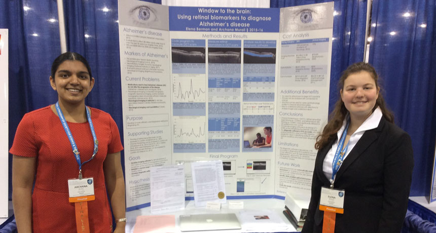
Archana Murali (left) and Elena Berman (right), present their eye study at the Intel International Science and Engineering Fair.
E. Nguyen/SSP
PHOENIX, Ariz. — The eyes of people with Alzheimer’s disease are, in some aspects, distinctly different from those of folks without this malady. That’s the finding of two teens. The changes they identified could offer reliable evidence of the disease in living people, the girls propose. More importantly, early signs of these changes might allow treatment for the disease before major symptoms occur. Right now, doctors and patients get no such early warning.
Archana Murali and Elena Berman presented their inventions here, this week, at the 2016 Intel International Science and Engineering Fair. Both 16-year-olds are 11th-graders at Breck School in Golden Valley, Minn. This year’s ISEF competition brought together more than 1,600 students from over 70 countries. Created by Society for Science & the Public, ISEF is sponsored by Intel. (SSP also publishes Science News for Students.)
Alzheimer’s disease is a horrible scourge. It causes confusion, mood changes and problems with memory. This brain disease cannot be prevented or cured. It also is the sixth leading cause of U.S. deaths. About 5.3 million people in the United States currently have Alzheimer’s disease. But they are not the only people to suffer from their illness. Family members and caregivers also face huge emotional and financial burdens, note Archana and Elena.
There are few sure-fire tests for Alzheimer’s disease. Blood tests won’t reveal it. Some genes can boost the risk of developing this disease. But people with those genes won’t always develop symptoms, Archana and Elena say. And Alzheimer’s symptoms may be confused with those of several other diseases. The only sure way to confirm an Alzheimer’s diagnosis is to look at samples of brain tissue after a victim has died.
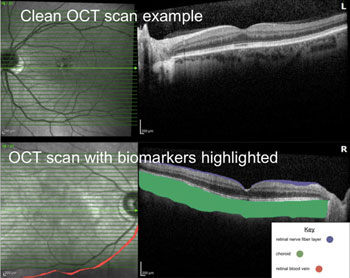
For instance, the teens measured the thickness of a nerve-rich layer in the retina (REH-tih-nah). That’s the light-sensitive layer inside the back of the eye. This fiber layer in Alzheimer’s patients ranged between 20 and 25 micrometers. That’s smaller than the 30 to 34 micrometers typical of the healthy volunteers.
The teens also measured the thickness of the choroid (KOR-oid), a blood-vessel–rich layer behind the retina. In healthy people, it ranged from 180 to 218 micrometers thick. But in Alzheimer’s patients, it typically ranged between 213 and 256 micrometers. Although those ranges overlap slightly, on average the Alzheimer’s patients had choroids that were much thicker, Archana and Elena note.
Finally, the teens measured the diameter of a major vein inside the retina. In Alzheimer’s patients, it typically measured between 83 and 94 micrometers. But in people without Alzheimer’s, the vein normally ranged between 131 and 134 micrometers.
At this point, it’s not clear what’s causing these eye changes, the teens say. Plus, it’s not clear when these changes might first emerge in an Alzheimer’s patient. Additional research might answer these questions. If Alzheimer’s disease indeed is behind these newfound eye changes — and they show up early in the development of this disease — then they could become an easy, low-cost means of diagnosing the disease.
Today, brain scans are only 75 percent accurate in diagnosing Alzheimer’s disease, Archana and Elena note. Based on their early analysis, the new technique appears to be more than 95 percent accurate. It also would be far less expensive, the teens say. While the cost of a brain scan is around $2,600, eye doctors can take an image of a patient’s eyes for about $100. So, if proven, the teens’ new technique could easily become part of a routine eye exam.
Power Words
(for more about Power Words, click here)
Alzheimer’s disease An incurable brain disease that can cause confusion, mood changes and problems with memory, language, behavior and problem solving. No cause or cure is known.
choroid The blood-vessel–rich layer of tissue that lies immediately behind the retina, the light-gathering layer of the human eye.
diagnose To analyze clues or symptoms in the search for their cause. The conclusion usually results in a diagnosis — identification of the causal problem or disease.
gene (adj. genetic) A segment of DNA that codes, or holds instructions, for producing a protein. Offspring inherit genes from their parents. Genes influence how an organism looks and behaves.
retina A layer at the back of the eyeball containing cells that are sensitive to light and that trigger nerve impulses that travel along the optic nerve to the brain, where a visual image is formed.
vein Part of the body’s circulation system, these tubes usually carrying blood toward the heart.


