Scientists mapped every nerve cell in this insect brain
Charting a baby fruit fly’s nerve cells and their connections took 12 years
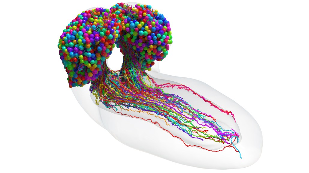
This diagram shows every nerve cell (multicolor) in a baby fruit fly brain. Each sphere represents a cell body. The elongated tails show the branches that send and receive information between neurons. The connections between the cells, known as synapses, aren’t visible here.
Michael Winding
Share this:
- Share via email (Opens in new window) Email
- Share on Facebook (Opens in new window) Facebook
- Share on X (Opens in new window) X
- Share on Pinterest (Opens in new window) Pinterest
- Share on Reddit (Opens in new window) Reddit
- Share to Google Classroom (Opens in new window) Google Classroom
- Print (Opens in new window) Print
The wiring of one insect’s brain is now pretty much mapped out.
Brains are full of nerve cells, or neurons. These neurons constantly send messages to one another. That’s how the brain functions, allowing animals to perform activities such as walking, breathing and thinking.
Human brains, which are estimated to have more than 80 billion neurons, are very hard to study. But many animals have smaller brains with fewer neurons. That makes them useful for learning how brains work.
For the first time, scientists have mapped all the neurons in a baby fruit fly brain. The researchers also found how the cells connect with one another. Such a map of neural connections is called a connectome. This is now the most complex diagram of a whole brain’s wiring ever created. Researchers described the work in the March 10 Science.
More complex brains
Until now, just three animals have had their brains diagrammed to this level of detail. One was a tube-shaped marine critter called a sea squirt. The two others were types of worms. But those creatures have at most about 1,500 neurons in their entire body. The scientists who did the new study wanted to understand much more complex animals, whose brains alone contain thousands of neurons.
Fruit flies (Drosophila melanogaster) fit the bill. Their brains more closely resemble people’s brains than the brains of a sea squirt or worm. Fruit flies also share a wide range of behaviors with humans. For instance, they can combine information from multiple senses, like sight and touch. They can also learn. This made the fruit fly brain an exciting new challenge in neural mapping.
The researchers decided to look at baby fruit fly brains, in particular. Baby fruit flies, called larva, can do nearly everything that adult flies can, except for some things like flying and mating. But the younger creatures have fewer and smaller neurons than adults, says Albert Cardona. This makes collecting data much faster, he says. A neuroscientist, Cardona works at the MRC Laboratory of Molecular Biology. That’s in Cambridge, England.
Mapping neurons
The team started sketching out the larval fruit fly brain 12 years ago, says Marta Zlatic. She’s also a neuroscientist at the MRC Laboratory of Molecular Biology.
First, Zlatic, Cardona and their colleagues captured images of the entire larval fruit fly brain. They did this using a powerful tool for zooming in on tiny things called an electron microscope. The researchers stitched those images together using a computer. Then they manually traced each neuron to create a 3-D virtual model of all the cells.
That model helped the team spot the connections where information gets passed between the neurons. These are called synapses. The researchers could even tell which cell sends information and which one receives information at each synapse.
In all, the 3-D model revealed more than 3,000 neurons and about 550,000 connections.
Patterns of connection
In the brain, collections of neurons called circuits work together to process information. So, the team explored how lots of neurons are connected to form circuits in fruit flies. The researchers examined neurons directly linked to one another. They also looked at distantly connected neurons that are not directly linked — kind of like finding connections between distant relatives in a family tree. Neural connections at this broader scale form what is called a connectivity pattern.
The connectivity patterns found in the fruit fly brain revealed 93 different types of neurons. Cells within each type share a similar pattern of connections to other cells. These groupings matched up with other features shared by the neurons, including shape and function.
Some neurons were extremely well-connected. They were directly linked with 20 or more other neurons. Nearly 75 percent of these neuron “hubs” were tied to the brain’s learning center — which goes to show how important learning is for these animals.
Although this work took 12 years, technology has vastly improved since the project began, Zlatic says. It would not take nearly as long if repeated today. But this whole-brain map may serve as a blueprint for other scientists studying brain circuitry, Zlatic says. “Now, we have a reference map.”


