Neutrons are unveiling hidden secrets of fossils and artifacts
Images made with these particles have revealed details of dinosaur bones, mummies and more

This artist’s illustration imagines a beam of neutrons uncovering a hidden bone — part of an ancient fossil embedded in rock.
Natasha Mutch/SayoStudio
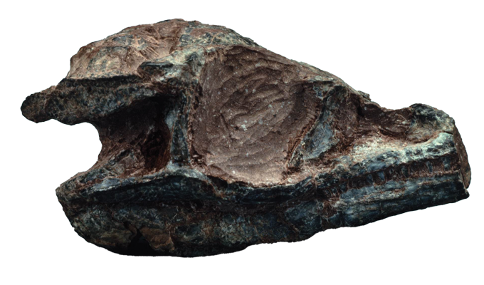
Some 100 million years ago, a crocodile ate a dinosaur for dinner. The croc died soon after, still stuffed with its dino victim. Later, its remains became encased in stone. This recently unearthed fossil has offered scientists a unique snapshot of life from the Cretaceous Period, when the croc prowled what is now Australia.
But the story of this crocodile’s last meal almost never came to light.
When Matt White first received the rock holding this fossil, his team scanned it with X-rays. White is a paleontologist at the University of New England in Armidale, Australia. Putting together multiple X-ray images of an object can create a 3-D map of its interior. White’s group hoped to use these scans to pick out each bone in the fossil without having to remove them from the rock.
And those scans did reveal much of the fossil. But iron-rich stone surrounding some of the bones blocked out X-rays. Needing some other way to peer through the rock, they sent the fossil to Joseph Bevitt. He’s a chemist at the Australian Centre for Neutron Scattering in Sydney.
Bevitt uses subatomic particles called neutrons to image ancient objects. His neutron scans uncovered the same croc bones that White’s team had seen. But they also revealed one bone that looked like a dinosaur leg. It was where the crocodile’s stomach would have been.
“When I saw the neutron result and the little dino femur, I was shaking with shock,” Bevitt says. He was “both in awe and doubt” about what he’d found. But years of analysis — plus more X-ray and neutron scans — confirmed that first glimpse. Those neutrons revealed the chewed-up remains of a never-before-seen species of dinosaur. It was inside the croc’s belly.
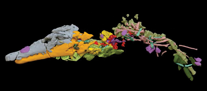
The discovery earned that crocodile — Confractosuchus sauroktonos — the second part of its name. Sauroktonos means “lizard killer.”
White, Bevitt and their colleagues shared their finding last year in Gondwana Research.
Neutrons have long been used to peer inside manufacturing and military equipment. In fact, people have used neutrons for imaging since shortly after the neutron was discovered in 1932. But these particles have only recently started giving scientists stunning views inside fossils and ancient objects.
Look, don’t touch
There was a time when studying fossils and artifacts required damaging or destroying them. Mummified remains were dissected. Sealed containers were cracked open. Fossils were pried loose from rock. Luckily, X-rays offer a way to inspect precious samples without ruining them.
X-rays are high-energy light waves. They interact with electrically charged particles, such as the electrons inside atoms. This allows X-rays to create images of the insides of objects. Here’s how it works.
Say a doctor wants to take an X-ray of someone’s leg to see if it’s broken. A machine shines a beam of X-rays through the patient’s leg. Those light waves get scattered or absorbed by the electrons in atoms that make up the leg.
Dense tissues, like bones, are packed with lots of electrons. That makes it difficult for X-rays to pass through these tissues. As a result, dense body parts — like bones — stand out in X-ray images. Skin, muscle and other soft tissues are much less dense. Since they have fewer electrons that can scatter or absorb X-rays, this radiation easily zips through the soft tissue. That’s why soft tissues essentially disappear in X-ray images.
X-rays have offered views into artifacts since this type of light was discovered in 1895. But in the 1970s, X-rays became the standard approach to studying fossils and artifacts.
They’ve recently revealed the insides of mummified animals from ancient Egypt. They also unveiled the brain cavity in a 20-million-year-old monkey skull. The machines used for this research are basically the same as the X-ray machines that doctors use.
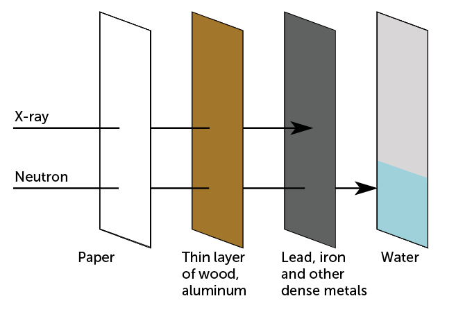
For all that X-rays have revealed about the past, they still have some drawbacks. X-rays can’t pass through super dense materials, such as lead or thick layers of other metals. So they can’t help researchers see inside such materials. On the flip side, objects made of low-density material, such as soft tissue, are invisible to X-rays.
Neutrons can fill in the picture.
Scatter!
Neutrons have no electric charge, so they ignore the electrons in atoms. So neutrons pass by electrons until they hit the cores, or nuclei, of atoms. Neutrons can bounce off an atom’s nucleus or be absorbed inside the atom.
These interactions are more complex than the ones between X-rays and electrons. How neutrons interact with atoms depends on how fast the neutrons are moving. It also depends on complex rules of quantum physics. (Quantum physics is the weird, twisty math that governs very, very, very small things.)
For instance, neutrons easily pass through lead, iron, copper and other metals that block X-rays. But neutrons interact strongly with some low-density materials through which X-rays easily zip. Lithium and boron are two examples. Another is water.
“Water to neutrons is like lead for X-rays,” Bevitt says. Why? Full of hydrogen atoms, water is like kryptonite for neutrons. It blocks them. Too much hydrogen-rich material can hide details from neutron beams.
But remember how a bone stands out on an X-ray image because it scatters or absorbs so many X-rays? Hydrogen can likewise make some features stand out in neutron images.
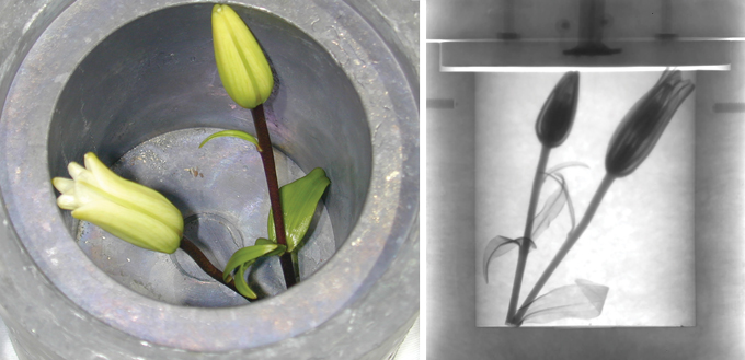
Jacob LaManna works at the National Institute of Standards and Technology, or NIST, in Gaithersburg, Md. This physicist likes to show the difference between neutron and X-ray imaging with flowers. He uses both techniques to image Asiatic lilies that he’s tucked inside a thick lead container.
“The neutrons can go right through the lead,” LaManna says. “Then you can see basically all the water [in the] vascular structure of the flowers.” But X-rays, blocked by lead, would show nothing but the opaque outer surface of the container.
Neutrons’ ability to glide through dense materials that block X-rays is their imaging superpower. It has made neutrons useful in the testing of cars and planes. The particles can reveal the flow of hydrogen-rich oil inside engine blocks. They can also expose flaws in metal castings.
Since the 1970s, U.S. national labs have relied on neutron imaging to develop and maintain nuclear weapons. Those neutrons can map the insides of dense bomb parts. They also can be used to study hydrogen-rich explosives inside nuclear warheads.
At NIST, LaManna heads a facility that can run X-ray and neutron scans at the same time. These dual views provide different insights into things that contain multiple materials. Hydrogen fuel cells, for example. Or building materials. Or soil samples. Such complex mixes of materials would be difficult to study with only X-rays or only neutrons.
Now, a growing number of paleontologists and archaeologists are taking advantage of this tech.
“We are really the new kids on the block,” Bevitt says of neutron-imaging experts. So far, neutron scans have helped study the fabric swaddling cat mummies without unwrapping them. Neutrons have also uncovered the most ancient vertebrate heart ever found. It was in a 380-million-year-old fish. Neutron beams have even helped experts spot fake ancient artifacts by finding signs of recently applied glues.

Rewards and risks
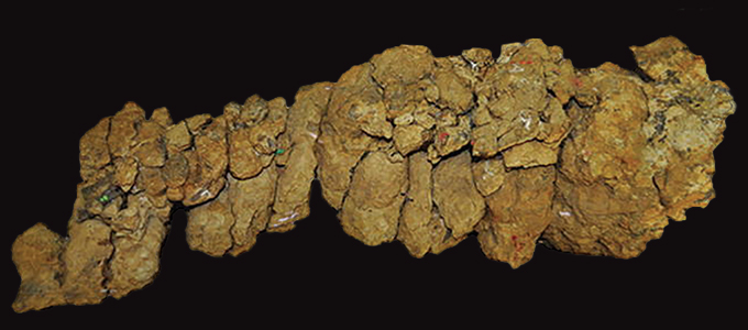
James Clark places a pair of fossilized crocodile skulls on the table in his basement lab. A paleontologist, he works at George Washington University in Washington, D.C. Clark’s 165-million-year-old croc fossils are dwarfed by a nearby modern alligator skull. That skull is about as long as my forearm. The fossilized croc skulls are only slightly bigger than my thumb tip.
Clark collected the fragile skulls in Mexico about 40 years ago. They’re embedded in hardened blobs of sediment with just a few bones and teeth peeking through. At first glance, the specimens look like wads of chewed gum. But they’re made of gritty, iron-rich mudstone.
“If you try to X-ray that, you basically end up with … these bright sparkles from all the iron,” LaManna says. The result are blurs and streaks. They mask the bones inside.
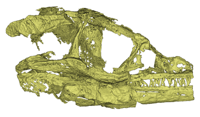
Clark could have hired someone to clean away the sediment around the delicate bones. But that’s a slow and costly job. Plus, he notes, it can harm the specimen. It wasn’t until 2019 that neutron imaging finally offered a good look at the hidden bones.
Iron is essentially transparent to neutrons, LaManna says. So “it’s much easier to basically isolate just the fossil” with neutron scans than with X-rays. NIST’s neutron scans revealed the intricate details of the tiny bones.
In the case of Clark’s crocodiles, it was the material around the objects that presented a problem for X-rays. But in other cases, the object itself can be the issue. Tissues, fibers, wood and other low-density materials can be difficult to spot using X-rays. And metals within an object can block other features from view. Both challenges plague researchers who are studying antiquities.
Digging into a dagger’s details
Ariel O’Connor is one of those researchers. An art conservator, she works at the Smithsonian National Museum of Asian Art. It’s in Washington, D.C. O’Connor was curious how some 3,000-year-old dagger-axes in the museum’s collection were put together.
These ceremonial weapons came from China’s Shang dynasty. They feature jade blades and turquoise-encrusted bronze handles. They also sport intricate blue-green patterns of gemstones sunk into the metal.
X-rays can’t tease apart the stone, metal, fibers and other materials that may be within the dagger-axes. Neutron imaging could help. But it also comes with a risk. Neutron beams make things radioactive. It’s not always clear how radioactive a sample will become. But materials often turn more radioactive than is safe for people to handle — or even view in a museum. That radioactivity can last days to weeks after a neutron scan.
The dagger-axes contain material that was “basically just completely dug up from the ground,” LaManna says. “It can have all sorts of stuff in it that you might not necessarily expect.” That made it hard to predict how radioactive scanning would leave the weapons.
O’Connor’s team decided to do a test.

They made a crude replica of an ancient dagger-axe. They used jade from Wyoming in place of the ancient Chinese jade. Brass from a door mimicked the bronze handle. The researchers even used some silk thread similar to the type that holds together some Shang dynasty dagger-axes. LaManna then scanned this dagger with X-rays and neutrons at NIST.
As expected, the brass completely blocked the X-rays. This hid features of the replica from view. But the neutron beam revealed key details, including jade inside the brass handle — even single silk threads.
The replica didn’t show any significant radioactivity nine days later. That’s normal, Bevitt says. Most samples are safe to send back to labs and museums within a few weeks. But it’s unclear just how chemically similar the replica was to the antique dagger-axes. So O’Connor is not yet ready to risk scanning the actual artifacts.
“I am entrusted with the preservation and safety of these remarkable 3,000-year-old objects,” O’Connor says. And if there’s any chance that neutron scans might alter them or prevent them from being displayed? “We will look for other options.”
A new window to the past
Despite the growing popularity of neutron scans, X-rays remain the go-to choice for many researchers. X-rays can uncover small details with no lingering radioactivity. What’s more, X-ray machines are widely available and small enough to fit in most labs and museum research spaces.
Right now, there are only a few dozen neutron-imaging facilities on the planet. Neutrons from those scans come from particle accelerators and nuclear reactors. Such machines are therefore large and quite costly. They also require a lot of oversight to work safely.
LaManna thinks there’s another major roadblock to more scientists using neutron imaging: They simply aren’t aware of what it can do. “I try to recruit as broad a range of users as I can” to submit fossils and artifacts for imaging at NIST, he says. He’s sees it as “getting the correct people interested.”
In the last decade, Bevitt has sung the praises of neutron imaging globally through lectures and outreach efforts. Many researchers in his home country of Australia have already embraced the tech. They bring objects to Bevitt for neutron scanning, just like White’s team brought their dino-eating croc fossil.
“Basically, in Australia, when a new dinosaur is discovered,” Bevitt says, “the first thing that happens is it comes to our lab.”


