Mummies share their secrets
Technology helps scientists understand how the dead once lived
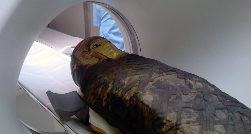
CT scans at the Field Museum in Chicago helped confirm the age and sex of seven Egyptian and three Peruvian mummies. A scan of this Egyptian mummy revealed it to be a woman about 40 years old.
The Field Museum, 2011
One afternoon, Ron Beckett, a professor at Quinnipiac University in Hamden, Conn., left his lab, taking some research home with him. The university labs were being remodeled, and security was a question. Carefully assisting his elderly passenger into the back seat, Beckett buckled her in for safety. As the two traveled to Beckett’s home, the passenger sat very still and didn’t make conversation — because she’s a 1,000-year-old Peruvian mummy!
Mummies are alive with information, and scientists like Beckett are helping to unlock what these time travelers have to say. Cutting-edge technology such as CT, or CAT, scans and endoscopes are allowing scientists to see not just what’s underneath the wrappings but also what’s inside a mummy’s body.

Mummies have been discovered all over the world, including Europe, Asia, Africa, Oceania and South America. Most people think of a mummy as a body wrapped in yards and yards of linen. But a mummy doesn’t have to be wrapped. Technically, any preserved body with some hair, skin and muscles is a mummy.
Mummies can be human-made. Ancient Egyptians are probably the most famous mummy makers. They dried out bodies with salts, then removed the internal organs that would decay and placed them in containers called canopic jars. Afterward, the bodies were wrapped in long swaths of linen.
Ancient Peruvians preserved their dead as well. People of Papua New Guinea mummified some of their dead through a smoking process — much as sausages and other meats are preserved today.
When conditions are just right, nature can mummify bodies. Preserved people have been found in the spongy peat bogs of northern Europe. Dry caves, deserts and frozen temperatures have also preserved people and animals that lived hundreds — even thousands — of years ago. This has allowed modern scientists to “meet” these ancients.
Technology helps mummies talk
While on assignment for National Geographic in 2008 and again in 2010, Beckett traveled to Papua New Guinea. He was there to study the mummy of Moimango, the father of a chief of the Anga tribe. Reaching this remote mummy proved challenging. For instance, Beckett crossed one treacherous bridge: a single log about 10 inches across and slippery from a persistent tropical downpour. Ever so slowly, in the dark and with no safety ropes, Beckett used it to cross a raging river.

Moimango, like other mummified chiefs and clan members before him, normally sits in a chair atop a 1,000-foot-high cliff overlooking the village of Koke (KO’ kay). Villagers recently carried Moimango down. Covered in a layer of clay, Moimango’s dried remains had a bright reddish color. But he was in very bad condition after 50 years out in the open. Lichens were growing on his toes and fingers, and some of those digits were barely attached. Facial bones lay bare and some skin had peeled away from his face.
Beckett brought along an especially helpful tool called an endoscope to study Moimango. An endoscope is a tiny camera about as big as a standard pencil eraser that can be snaked into a small opening on the body. Such a tool lets scientists see things that would otherwise be invisible. Beckett used his endoscope to investigate Moimango’s remaining organs and discovered this mummy had slight gum disease but teeth in good condition. The scope also showed nesting materials from a tiny rodent, perhaps a field mouse. Moimango’s brain was gone, but in its place Beckett found several wasp’s nests and a few very active wasps.
Recently, Beckett used X-rays to solve a mystery surrounding a 4,000-year-old Egyptian mummy. Pa-Ib (pronounced pie eeb) is part of the collection at the Barnum Museum in Bridgeport, Conn. Not much was known about this mummy. Ancient Egyptian writing on the coffin suggested this ancient had been male. And legend had it that the body hosted another mummy — of a bird. Beckett puzzled over how he might confirm these suspicions without unwrapping the mummy.
He decided to investigate using high-tech lab instruments.
Along with his Quinnipiac colleague Gerald Conlogue, Beckett carefully packed the mummy in acid-free paper and plastic bubble-wrap material. Then they placed Pa-Ib in a special container to protect the fragile remains during transport. Escorted by police, the mummy rode to the lab in a black sport utility vehicle.
In their lab, the scientists peered inside Pa-Ib using a machine called a computerized tomography — or CT — scanner. The device resembles a giant doughnut standing on end. As an object moves through the doughnut hole, the scanner takes hundreds of X-rays from a range of different angles. Then a computer processes the information to reconstruct a three-dimensional image of the object.
This scan revealed Pa-Ib had been a woman, not a man, about 5 feet 3 inches tall. Her stomach contained several unusual packets. To evaluate whether one might be a bird, Becket brought a recently mummified bird to scan and compare.
The scientist had found a dead falcon near his Arizona home and had used baking soda, table salt and linen to mummify it. A CT comparison of it and the packets inside Pa-Ib ruled out the presence of any bird inside the ancient Egyptian remains. So what’s inside her packets remains a mystery.
Gregory Thomas at the University of California, Irvine, also probes mummies with CT scanners. In his case, he has been studying the heart and blood vessels in 52 Egyptian mummies at the National Museum of Antiquities in Cairo.
Those X-ray reconstructions revealed several of the mummified individuals had suffered from heart disease. Although this is the leading cause of death in the United States today, scientists had long assumed this was a modern disease. But scans of the 3,500-year-old mummified remains of Princess Ahmose-Meryet-Amon now point to her being the oldest known case of heart disease.
“We are more similar to these ancient people than we thought,” concludes Thomas.
X-ray surprises
The Field Museum in Chicago is using CT scans to learn more about mummies in its collection. These X-ray reconstructions helped confirm the age and gender of seven Egyptian and three Peruvian mummies along with details on the contents and construction of their coffins.
The mummies were placed into a loaned scanner that was inside a trailer parked just outside the museum. Each “patient” was carefully wheeled on a medical gurney down hallways and up ramps to the waiting machine. To keep possible dirt, rain and bird droppings off the mummies, protective cloth covered them along the way.
Adding to the challenge, these mummies were longer than the scanner bed, so each individual had to get two separate scans— head to mid-thigh, and pelvis to feet.
“It was a challenge to keep the humidity and temperature inside the trailer at levels that enabled the machine and computer to work,” remembers J.P. Brown, who works at the Field Museum. “Once the mummies were inside, the scientists had to leave the trailer so their body heat and breath wouldn’t add to the heat and humidity.”
One Egyptian mummy looked great from the outside. But the scans turned up that this particular set of remains had no hips, arms or torso.
“It was a bit of a shocker, and our first reaction was the scanner had broken,” recalls Brown.
Another mummy scan showed tiny plugs, possibly made of wax, that had been placed in the nostrils to help keep the nose’s shape after it had been broken during mummification.
CT scans also offered clues to what ancient peoples ate. This past summer, scientists at the South Tyrol Museum of Archaeology in Bolzano, Italy, announced that they had studied the stomach contents of Ötzi (UHT-zee) and learned what his last meal was. Ötzi, a 5,000-year-old mummy, was discovered by hikers in 1991. The frozen remains had been preserved in glacier ice high in the Alps, along the Austrian-Italian border.
Scientists had searched for Ötzi’s stomach but couldn’t find it. A recent CT scan located it in his upper torso. Ötzi was found facedown, leaning over a rock, and scientists believe this position may have caused his stomach to slip upward. They also think that organ shrinkage might have caused many of his innards to shift their positions.
Researchers were surprised to find Ötzi’s stomach completely full. It contained yellowish and brownish materials along with pieces of grain and meat. The scientists removed a small sample and tested its DNA. It now appears Ötzi had feasted on an Alpine ibex, a type of mountain goat that lived in the region, approximately 30 minutes before his death.
There’s a saying that “Dead men tell no tales.” But thanks to science, this is no longer true. Technology is giving mummies a voice.
Power Words
mummy: A body preserved by natural processes or human technology, with some skin and organs remaining.
canopic jars: Containers used by ancient Egyptians to store the organs of dead people.
embalming: A process that preserves a dead body by preventing it from decaying.
peat bog: A soggy, brown, soil-like material made from decaying plant matter.


